


Consultation & OPD Timings
Meet with Dr. Mandeep Singh Malhotra at CK Birla Hospital
For AppointmentLeading the Future of Breast Cancer Surgery: Dr. Mandeep Singh Malhotra Completes 50 Robotic Nipple-Sparing Procedures
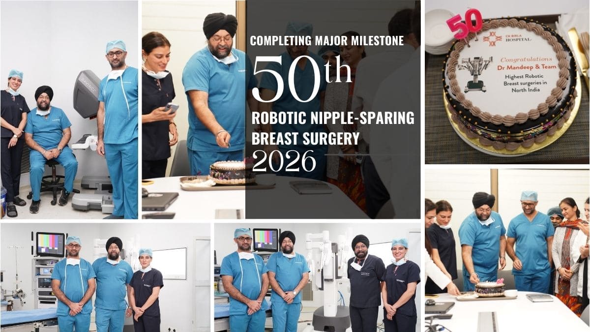
Dr. Mandeep Singh Malhotra has achieved a significant clinical milestone by successfully completing his 50th robotic nipple-sparing breast surgery, marking a historic first for North India. With this achievement, he becomes the first surgical oncologist in the region to reach this benchmark using robotic-assisted technology for nipple-sparing breast surgery.
Reaching this milestone reflects not only surgical expertise but also a deep commitment to innovation, patient-centered care, and continuously raising standards in oncoplastic and robotic breast surgery. It stands as a testament to how technology, when combined with experience and vision, can redefine outcomes for patients.
Dr. Mandeep Singh Malhotra-Surgical and Molecular oncologist in Delhi NCR India

MBBS, MS, Surgical Oncology Residency(AIIMS), Fellowship in Head Neck Oncology(MSCC-NH), Fellowship in Breast Oncoplasty(St Andrews UK), Robotic Certification for Trans Oral Surgery,(UPENN USA), Hyperbaric Medical Technology Certification(South Carolina, USA), Molecular Oncology Certification(RGCC Global College,Greece)
A Surgical Oncologist with sub-specialization in Head Neck & Breast Oncology and more than 20 years of patient treatment experience, Dr. Mandeep is currently working with CK Birla Hospital,Punjabi Bagh,New Delhi as Director-Surgical Oncology and has previously also worked with Fortis Hospital, Vasant Kunj, as Sr. Consultant & Head of Department – Head Neck & Breast Oncoplasty.
He is amongst the Most Experienced Surgeons in India, in treating and operating Advanced or Recurrent Oral (Mouth), Oropharynx (Throat), Larynx (Voice Box), Thyroid, Orbital and Maxillary (Upper Jaw Bone) Cancers.

Breast and Head & Neck Cancer Care Treatment!
Providing world class cancer treatment in India.
Breast Cancer
Breast cancer occurs when some breast cells begin to grow abnormally.lifestyle and environmental factors that may increase your risk of breast cancer.
Read MoreHead and Neck Cancer
Head cancer is site-specific, which includes oral cavity, pharynx, larynx, paranasal sinuses and nasal cavity, and salivary glands.
Read MoreThyroid Cancer
According to an article published in JCO (Journal of Clinical Oncology) in 2018, Thyroid Cancer has risen in India particularly among the younger population..
Read MoreBook an Appointment with Dr Mandeep?
Dr Mandeep Singh Malhotra is one of the best surgical oncologist in India. Presently working as the Head Oncologist at CK Birla Hospital, West Punjabi Bagh, Delhi.
Our Locations
Plot number 219p, HCAH Transition Care Centre, 226, Block N, Urban Estate, Sector 51, Gurugram, India, Haryana
OPD 11, 57/41, CK Birla Hospital, Rd Number 41, West Punjabi Bagh, New Delhi, Delhi 110026
Call Now
+91 9971474985
Medharbour Multispeciality Hospital, Gurugram, Plot Number 222, Sector 51,
Gurugram, 122003
Call Now
+91 8471002000